Discover TD-NIRS
Learn how Time Domain Near-infrared Spectroscopy can add a new dimension to your research
How does it works?
Time Domain Near-infrared Spectroscopy (TD-NIRS) is a non-invasive optical technique that uses low-power laser light to measure various biomarkers in humans, like functional activations in our brain or hemodynamics in our muscles.
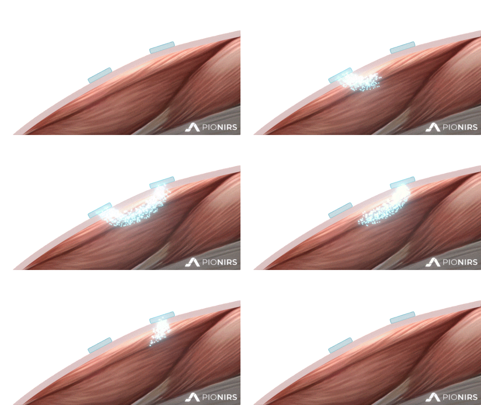
Propagation of short light pulses (with a duration lower than 200 ps) in the tissue, hindered by multiple absorption and scattering events, deeply modifies their temporal shape which is eventually reconstructed (photon by photon) by a very sensitive photodetector.
A key feature of TD-NIRS is its capability to add a new dimension to classical NIRS measurements, thanks to recording of the time that each individual photon takes during its trip through the sample.
Indeed, the strong relationship between penetration depth of photons and their arrival time at the photodetector, makes it possible to select the position in the tissue at which we want to retrieve the information.
This is a crucial advantage to better estimate deep functional variations, while neglecting the contribution of unwanted superficial and systemic changes.
Here at PIONIRS, we overcame the bulkiness and complexity of old-generation TD-NIRS systems and are now able to provide an innovative, compact and easy to use new generation of devices for your biomedical research.
Time Domain Near-infrared Spectroscopy (TD-NIRS) is a non-invasive optical technique that uses low-power laser light to measure various biomarkers in humans, like functional activations in our brain or hemodynamics in our muscles.
How does it work?
Propagation of short light pulses (with a duration lower than 200 ps) in the tissue, hindered by multiple absorption and scattering events, deeply modifies their temporal shape which is eventually reconstructed (photon by photon) by a very sensitive photodetector.
A key feature of TD-NIRS is its capability to add a new dimension to classical NIRS measurements, thanks to recording of the time that each individual photon takes during its trip through the sample.
Indeed, the strong relationship between penetration depth of photons and their arrival time at the photodetector, makes it possible to select the position in the tissue at which we want to retrieve the information.
This is a crucial advantage to better estimate deep functional variations, while neglecting the contribution of unwanted superficial and systemic changes.
Here at PIONIRS, we overcame the bulkiness and complexity of old-generation TD-NIRS systems and are now able to provide an innovative, compact and easy to use new generation of devices for your biomedical research.
Advanced data analysis
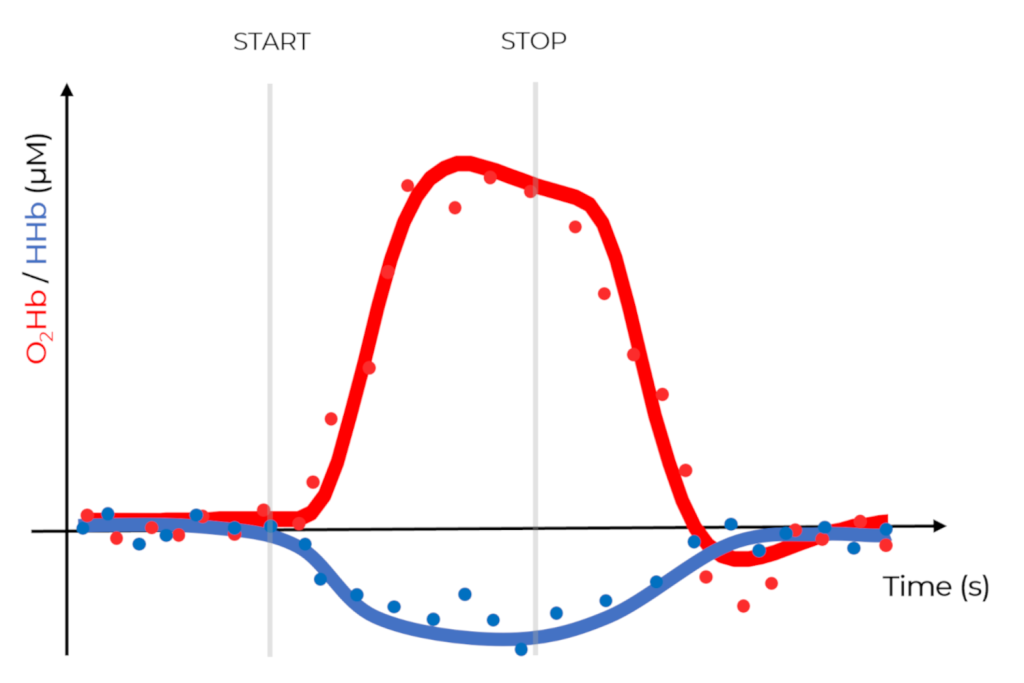
Exploiting data processing and dedicated light-diffusion physical models, it is finally possible to retrieve absorption and scattering coefficient of the tissue under investigation, that can lead us to the estimation of concentration of oxy- and deoxyhemoglobin.

Selected scientific literature:
Interested in having a more detailed description of what we do and how TD-NIRS could be useful for you? Take a look at these publications and get in touch with us!
A comprehensive review on TD-NIRS: features, advantages, hardware and applications.
–
NeuroImage
Vol. 85, Part 1, January 2014
Example of TD-NIRS applied to muscle fatigue assessment during isometric exercises.
–
Biomedical Optics Express
Vol. 11, Issue 12, December 2020
Our NIRSBOX device in a study on hemodynamics monitoring in free-walking exercises.
–
Neurophotonics
8(1), 015006, February 2021
